OPM:
Ocular perfusion maps from retinal fluorescent angiograms
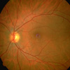
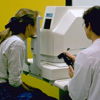
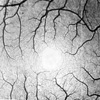
The ocular fundus (background of the eye, see fig. 1) offers the unique opportunity to visualize the blood vessel system in vivo. Scanning ophthalmoscopes such as the Rodenstock LSO® (fig. 2) deliver high–resolution dynamic image sequences at video frame rates which resolve vessels close to the size of the capillaries (fig. 3 foveal region).
Many eye diseases such as age–related macula degeneration (AMD) are correlated with irregularities in the metabolism with effects on the retinal blood flow. The perfusion of the bood vessel system is, moreover, affected by systemic diseases such as diabetes ors hypertension. These effects can also be observed on retinal blood vessels. Hence the evaluation of retinal perfusion would provide early information of the onset of those diseases.
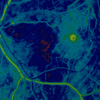
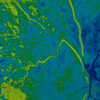
The perfusion of the retinal blood vessels can be observed dynamically by means of an intraveneous application of fluorescent agents (e.g. fluorescine, ICG), the so–called retina angiograms. By means of a sophisticated registration process the indispensable eye movements—even sudden large saccades— are compensated to sub–pixel accuracy, see motion picture sequence (the moving frame demonstrates the compensated movements). The delay of the local arrival of the contrast agent bolus indicates the local distribution of the blood velocity. Appropriate color coding schemes yield functional images which offer a quick interpretation of the
Calibration of above functional images by distributed local measurements of the blood flow velocity (e.g. by laser doppler velocimetry) yields a further improvement. To achieve a correct spacial correlation between images and local measurements the images have to morphed on a sphere model of the eyeball which results in so–called ocular perfusion maps.
Medical partner:
Department of Ophthalmology, Aachen University Hospital
Project period: 1991 to 2000
 1
1
 2
2
 3
3
 4
4
 5
5