MMCA:
multimodal cell analysis for early cancer detection and diagnosing
An ongoing project of the
Institute of Imaging and Computer Vision, RWTH Aachen University
At cellular level—the cytopathologic analysis of isolated cells and their nuclei—cancer can often be discovered earlier and sometimes be identified even more accurately, at lower costs and with less discomfort for the patient than at tissue level (histopathology) or at organ level (radiology, nuclear medicine). In particular computational approaches to quantitative analysis of cells and sub–cellular structures yield reliable diagnostic information beyond the traditional subjective visual inspection.
A further remarkable improvement of the diagnostic precision is obtained by a multimodal cell analysis (MMCA): MMCA exploits more exhaustively the information provided by both visible light and fluorescence microscopy by the fusion of diagnostic data for individual cells. To this end, the specific diagnostic information of complementary evaluation methods, i.e. different stainings and marker demonstrations applied to the cells, are accumulated cell–wise by a precise registration of the cells and their nuclei after successive re–staining processes. Thus, improved reliability and expressiveness of diagnoses is achieved on even lower numbers of cells.
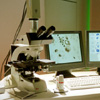
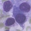
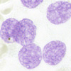
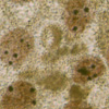
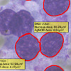
Figures at right show a protype mmca workstation (1), morphologically stained cells (2), DNA–stained cells (3), cells with AgNOR demonstration (4), and registered cells labeld with the quantitative results yielded from the three above stains (5). Click on thumbnail to blow up! >>
Medical partner:
Prof Dr med Alfred Böcking,
Institute for Cytopathology, Univ of Düsseldorf
Scientific partner:
Prof Dr Branko Palcic
British–Columbia Cancer Agency, Vancouver BC, Canada
Industrial partner:
Motic ltd, Xiamen, PR China
Project period: 2001 ongoing
Links:
Inst. of Imaging and Computer Vision, RWTH Aachen Univ., (project site)
Special site on cytopathologic cancer diagnostics
 1
1
 2
2
 3
3
 4
4
 5
5